Julie Prebel, Occidental College
(Published August 21, 2015)
In 1844 Eliza Farnham (1815-64) was appointed matron at the women’s prison at Sing Sing (Mount Pleasant State Prison) in New York, embarking on a four-year term during which she proved an especially controversial figure in the already contentious debates on prison and criminal reformation.1 Farnham’s beliefs about criminals, especially female criminals, and her methods for reforming them make her unique in this cultural history. She believed in the possibility of rehabilitation, contrary to predominant cultural and scientific views, and pursued the goal not through the conventional theological approach of religious conversion but through the use of phrenology. Although dismissed today as a pseudoscientific fad grounded in unsound theories, and even considered fallacious by many in the nineteenth-century, phrenology attracted the interest of reformers, like Farnham, who advocated a scientific means of identifying and potentially altering criminal and other behavioral inclinations by detecting defective regions in the brain.
My interest in this article is not so much in tracing the history of phrenology, which is a well-trenched area of scholarship, or even in providing a detailed biography of Farnham’s role as a reformer, though certainly I touch on both points.2 Instead, I will focus on one aspect of Farnham’s contribution to the popularization of phrenology: a series of sketches and daguerreotypes she appended to a reprinted edition (1846) of a widely read text on crime and phrenology, Rationale of Crime and its Appropriate Treatment, by Marmaduke Sampson.3 Farnham’s series of cranial outlines of male and female criminals followed by nineteen daguerreotypes she commissioned from famed nineteenth-century photographer, Matthew Brady, functions as a register of her views on phrenology and criminal-prison reform. Just as importantly, however, Farnham’s images are “cultural and rhetorical forces” that perform a “rhetorical transaction,” in the sense Hill and Helmers describe, acting rhetorically on viewers-readers by attempting to influence their attitudes, opinions, and beliefs about the causes of and treatments for criminal behavior (21).
In this article, I read Farnham’s phrenological sketches and photos as emblematic of a distinctive moment in the scientific history, traced by Lisa Cartwright and others, of “surveillant looking and physiological analysis,” one means of diagnosing, classifying, and maintaining control over subjects deemed diseased, aberrant, or criminal (Cartwright 5). In this way, Farnham’s images are artifacts of a visual culture of scientific observation with contemporary relevance, as I explain below, part of a long history of techniques designed to “look” at and interpret human physiognomy and physiology. Farnham’s images can be understood as an early form of visual technology that purported a means to peer into the human body and psyche, and are tied to the historical development of “body analysis and surveillance in medicine and science” (Cartwright 3). In my reading of Farnham’s images, I understand them, as Cara Finnegan suggests in her work on the rhetorical aspects of visual culture, not merely as “products” consisting of “non-verbal or non-discursive features;” instead, I analyze them using a “critical and theoretical orientation” to view them as visual discourse (198). By employing “visual rhetoric as a mode of inquiry,” my aim is to underscore Finnegan’s distinction between analyzing “images in history” versus understanding visual “images as history” (198-99, my emphasis). In other words, Farnham’s phrenological images are not merely a way to illustrate or punctuate a certain historical moment, but instead contribute to a “rhetorical history of the visual,” comprising a continuum of visual methods that attempt, in particular, to render visible the inner workings of the mind (Finnegan 211).
In approaching these phrenological images as a rhetorical historian interested in the visual culture surrounding this archive and its broader implications, I connect Farnham’s nineteenth-century visual representations of heads to contemporary transformations in scientific visual technologies that extend the “medical gaze” into “domains that…had only been available through surgical or autopsical interventions” (Burri 39). Specifically, as I explore below, phrenological head sketches and photos are analogous to the forms of “digital body slices” produced by visualization technologies in medical sciences in the twentieth-century, and especially since the 1970s, such as ultrasound, computer tomography (CT), magnetic resonance imaging (MRI), positron emission tomography (PET), and digital X-rays (Burri 38). In the past decade especially, the functional MRI (fMRI) has been increasingly used by neuroscientists and psychologists to answer questions about which part of the brain is responsible for certain behaviors, feelings, inclinations, and decision making processes—not unlike the predictive promises of phrenology. As I argue below, Farnham’s images are early examples of such scientific visualization, and in both cases there is a pictorial turn that brings about epistemic changes in the scientific surveillance of the human body.
It may seem somewhat obvious to consider a practice that relied on visual narration a mode of visual rhetoric, yet notably historians and cultural critics have been slow to explore the role of visual elements in phrenological discourse. Allan Sekula has described the conjuncture of phrenology and photography in his work on the rise of photography in the nineteenth-century as a bourgeois mechanism for constructing and cataloging the criminal body, touching on the collaboration between Farnham and Brady as “the first sustained application of photography to the task of phrenological analysis” (Sekula 9). Sekula’s main interest is to read the concurrent proliferation of photography and phrenology as part of the “taxonomic ordering” that “contributed to the ideological hegemony of capitalism” in an era of heightened interest in constructing cultural typologies based on bodily forms and appearances (12-14). My focus here is no less ideological, especially as gender, class, and racial standards are encoded into Farnham’s interpretations of her image archive, as she ranks the phrenological subject’s mental capacities according to visually apparent physical characteristics.
An emphasis on the verbal-visual link evident in Farnham’s text further distinguishes my work from Sekula’s as grounded in rhetorical praxis. As I show below, Farnham uses both word and image with fluidity, as both act as “symbolic forms of representation” that “work” upon her readers-viewers (Hill and Helmers 2). Not only does Sekula neglect part of Farnham’s archive—the hand drawn sketches—in favor of establishing the Brady photographic connection, he also discounts Farnham’s verbal descriptions that accompany the images. In keeping with Maureen Daly Goggin’s claim that there is not a “hard line of distinction between rhetoric of the word and rhetoric of the image,” I show how Farnham’s sketches and photographs, accompanied by her verbal descriptions of them, engage the word-image dialectic central to rhetorical theory in provocative ways, especially in terms of ekphrastic representation (Goggin 87).4 Farnham’s contributions to Sampson’s treatise reveal such correspondences between written description and visual texts, as images and words “operate synergetically” and provide a rhetorical analytic frame for representing the criminal and degenerate (Goggin 88).
As Cartwright points out in her historical study of scientific visual culture, methods for surveying the human body, especially to uncover, diagnose, and treat certain pathologies, may have changed from the nineteenth-century with more skillful technologies but “surveillance of the body continues to be an important technique in medical practice,” most notably in neurology, which has scientific roots in phrenology (47). Phrenology (along with anthropometry and craniometry), as Cartwright notes, can be understood as “part of a broader set of typological systems” designed to provide empirical evidence of what the body’s surfaces might reveal; while it might be tempting to dismiss these nineteenth-century pseudosciences, this “visuality nonetheless survived its association with empiricism” and “has emerged at the center of a vast medical industry and subspecialty encompassing a plethora of postempirical imaging techniques” (51). Both phrenology and contemporary brain imaging are visualization techniques that allege a range of medical and social uses, from more benign identification of behaviors to determining criminal culpability. In connecting this visual rhetorical history in the sections below, I focus specifically on the phrenological readings and neuroimaging used to explain abnormalities and to assess criminal behaviors and propensities. Phrenology and brain imaging might be separated by the kairotic conditions of time and place, but they connect as sites of persuasion that encourage readers-viewers to practice new modes of seeing.
Phrenology as Visual Rhetoric
At its most basic, phrenology purported that the qualities of individual character originated in a system of distinct organs in the brain that housed the mental faculties controlling specific instinctive and behavioral traits, such as spirituality, benevolence, combativeness, or conscientiousness. Proponents of this science claimed that these human behaviors and propensities could be determined by a physical examination of the head, and that this topography of the skull provided an index of the corresponding identity beneath. As a diagnostic practice, phrenological science alleged the capability to identify and treat mental pathologies by locating defective traits in the skull’s indentations, while at the same time promising individuals the opportunity to affirm and enhance their personal and social potential by identifying and then developing latent aptitudes and dispositions.5
Expanding this theory of a relationship between internal and external, German anatomist and physiologist, Franz Joseph Gall, pioneered early research in neuropsychology by claiming to detect the various mental functions of the brain. Gall, considered the founder of phrenology, captured popular attention by claiming to show how individual psychological and behavioral differences were observable through the shape and irregularities of an individual’s skull. Gall’s work in the early decades of the nineteenth-century influenced developments in anthropology (especially craniology), psychology, and neurological sciences, and Farnham even went so far as to replace some of the religious instruction in Sing Sing with readings from Gall, believing in the persuasive promises of his “Science of Humanity” (Woman and Her Era 18).
Phrenologists in mid nineteenth-century America, such as Orson and Lorenzo Fowler, who helped popularize the science by linking it to an American patriotism interested in self-improvement and social change, claimed legitimacy for phrenology by tracing its roots to Gall’s practices of collecting, observing, and dissecting skulls. In their popular publications, such as The American Phrenological Journal and The Illustrated Self-Instructor in Phrenology and Physiology, the Fowler brothers persuaded their readers to become amateur phrenologists by guiding them through the steps of a diagnostic exam. Using both verbal text and illustrations, the Fowlers promised the means to master the interpretive codes that would unlock dormant mental faculties and enable personal growth and social mobility through their display of “Life Illustrated.”6
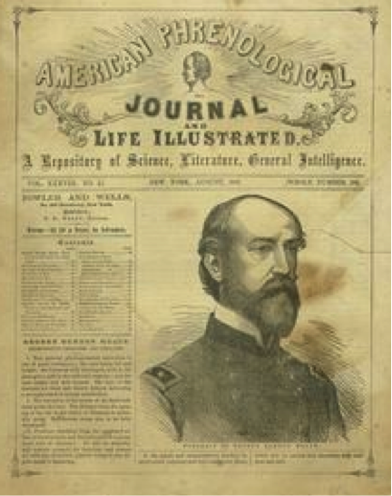
Figure 1: American Phrenological Journal
In the Self-Instructor, the Fowlers included a “phrenological diagram” showing thirty-seven numbered “organs” of the brain, which they intended as “illustrative” proof of their “very extensive professional observations and experiences.” They explained that their objectives were both descriptive and instructive: by using images or “engravings” they claimed to give their readers “at a glance...specific directions [about] how to perfect their characters” (vii, my emphasis). The interaction between the persuasive and the visual is thus very evident in these widely popular texts, as the Fowlers use drawings, tables, photographs, and figures to guide readers towards “cultivating, restraining, and rightly directing” their “mental faculties” (viii). The Fowlers encouraged their readers-viewers to engage with the visual and to associate these visual encounters with an increased self-knowledge and self-awareness.
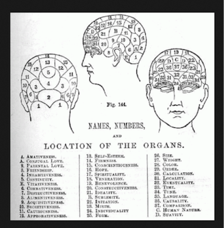
Figure 2: Numbered Phrenological Head Diagram
The Fowlers—along with other phrenologists of the era, Farnham included—used images in a way that showed a reliance on visual representation and encouraged visual literacy on the part of lay viewers. In his work examining the use of mechanical devices used to generate visual data and to chart physiological evidence by nineteenth-century scientists, which included a range of instruments from microscopes to photographic apparatus, Joel Snyder claims that the “visualizations” produced by these machines are forms of “graphic data” designed to act as “aids to the eye” of the viewer, enhancing their sense of sight and showing “things that cannot be detected by the ‘unaided eye’” (383). In one sense, phrenological images—and in some cases three-dimensional skulls of living and dead people—acted in precisely this way, as “graphic data” that made visible or displayed certain external realities otherwise overlooked by the seer. In another sense, though, the semiotic use of head images by phrenologists performed an important rhetorical function, as the study of these visual symbols—the data shown through head bumps—promised to “disclose specific signs of character” and thus give people the means “by which we may know ourselves and our fellow-men with certainty” (Illustrated Self-Instructor 11). In this interaction between the phrenologist, the image, and the viewer, there is the notion that scientific authority helps to corroborate human vision; if the viewer is not sure what to look for in the image or what they should “know” by observing it, the phrenologist will point them towards an interpretation. Constructing this rhetorical meaning of “self” and “other,” though, also depends largely on the participants—in this case the audience enlisted as armchair phrenologists able to scan human heads for signs of abnormalities and tendencies.
This “do-it-yourself” approach to the science of phrenology thus both authorized the viewer’s vision and encouraged rhetorical action; after all, phrenology was a scientific tool with the potential use-value to improve individuals and all of humanity. Self-knowledge and the ability to know others through the meaning attributed to phrenological signs also fit nicely into nineteenth-century cultural ideologies about personhood and self-improvement, especially when the qualities to be improved were more nonthreatening to any sense of social order. For example, despite promoting phrenology as a scientific tool to improve humanity, Gall, the Fowlers, and other phrenologists of the era viewed criminality as an inherent—and in many cases inheritable—mental illness, the features of which could be identified but not necessarily “cured.” They used phrenology to explain criminal behavior as a biological sickness, caused by defects in particular faculties of the mind, and these were theories that influenced criminal anthropology at the turn of the century—especially the work of Cesare Lombroso.7 While contemporary theories of crime explain criminal behavior in both biological and social terms, many of the current methods for detecting criminality remain grounded in similar phrenological concepts from the nineteenth-century, especially in the use of scientific images as evidence to correlate brain activity and behavior, as I will show in the sections below. Farnham’s work is a key transition point in this discourse, and her use of images in Sampson’s text shows her attempts to close the visual rhetorical gap between “seeing” and interpretive meaning.
Farnham’s Persuasion of the “Popular Mind”
In her introductory preface to Sampson’s text, Farnham articulates her intention to provide a scientific visual technique for observing and quantifying human behaviors, abilities, and characteristics, and that she had a rhetorical aim in mind, which was to “adapt” this subject of “Criminal Jurisprudence…to the purpose of interesting the popular mind” (Rationale xxxi). After thanking the Fowlers, “for aiding me in the selection of cases” and the photographer, Brady, who produced a “very accurate set of daguerreotypes,” Farnham guides her readers-viewers through a phrenological analysis of her selected criminals, primarily women, and what defects or propensities their heads reveal (xxxviii). Farnham’s sketches and photographs are artifacts created and arranged with a sense of what Sonja K. Foss calls “purposive production,” as images “generate[d]…for the purpose of communicating” with an audience, intentionally designed to guide her readers towards an understanding of how the development of certain organs of the brain lead somewhat inevitably to criminal acts and behaviors (Foss 304). While the drawings rely in part on Farnham’s verbal text to provide them with greater meaning, the images themselves also perform what Foss calls a “symbolic action” as somewhat “arbitrary” referents that invite a viewer’s rhetorical response. Farnham urges her audience to imbue these skull outlines with meaning as they scan these heads for the signs of degenerative propensities, though the simplicity and sparseness of the drawings seem to belie any obvious or “natural relationship” between the sign and its object (Foss 305).
For example, Farnham’s first cranial outline, Drawing No 1, would appear to have very little interpretive meaning on its own without Farnham’s verbal text to guide her readers-viewers towards the conclusion that “No 1 is the head of a very depraved person…with a very large development of the inferior propensities” (Rationale 8).

Figure 3: Farnham Cranial Outline No. 1
Just as the visual drawing depends on the accompanying verbal text, so, too, does Farnham’s lengthy description of No 1 tell readers what to look for in the image, as the outline serves as the visual proof of this criminal’s “rudeness and coarseness,” evidence of his “sanguine and lymphatic” temperament—all of which result in the diagnosis that the “circumstances of his crime are strongly indicative of his character” (Rationale 8). The cranial drawing of No 2 also confirms the apparent arbitrariness of the visual sign, as Farnham’s audience is encouraged to derive meaning from yet another simple outline, which ostensibly reveals this criminal’s “largely developed” organ of “destructiveness.” In claiming that this “drawing indicates an exceedingly imperfect development of the superior sentiments,” Farnham attempts to persuade her readers-viewers to see what might not otherwise be overtly evident by encouraging them to look for these qualities in the image (9). In many ways, these hand drawings are nearly indistinguishable from one another, suggesting that Farnham’s pictorial impulse lies in the ways she brandishes an easy-to-read map of these human heads. Through image and text, Farnham advances similar enthymematic arguments about all of these head drawings, asking readers-viewers to participate in their own persuasion by filling in certain unexpressed premises: in this case, these premises have to do with what a curve in the head sketch might mean, or what might be known about an individual’s characteristics because of their head shape.
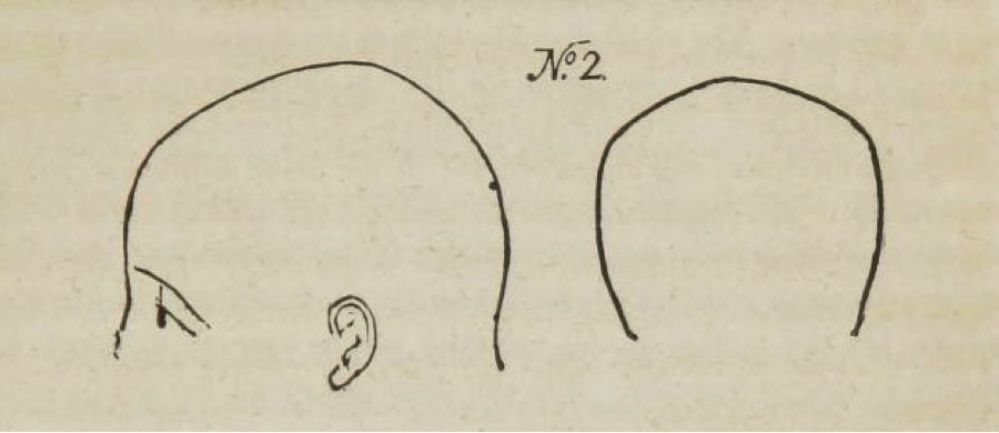
Figure 4: Farnham Cranial Outline No. 2
Farnham’s version of DIY phrenology is similar in many ways to the persuasive use of visual and verbal texts by the Fowlers and other phrenologists, as she attempts to convince people which human features and qualities should be observable by the human eye—and trains the public in terms of what to look for. Part of this training of the public to become amateur phrenologists is ideological, and so not quite as benign or objective as these scientific proofs might appear. The Fowlers, following the examples of other prominent phrenologists such as Gall and George Combe, frequently used their numerous illustrations and 3D displays of heads as evidence in their arguments for discernable differences between men and women and variations among races. As historian Carla Bittel notes in her work on phrenology in the early and mid decades of the nineteenth-century, “phrenology, at is core, was a taxonomy; men and women, ‘civilized and savage,’ were categorized, compared and contrasted” (109). The Fowlers, for example, drew such delineations especially in terms of racial hierarchies and argued (in Phrenology Proved, Illustrated, and Applied) that “men and women of European origins had ‘intellectual and moral superiority over all other races’” (Bittel 109). In looping back to the persuasive aspect of the phrenology practiced by the Fowlers and by Farnham, then, it is clear that along with training audiences to see differently they were also “instructing audiences on the hierarchies of nature,” which they believed to be evident in the visual scanning of skulls (Bittel 109).
Two of Farnham’s sketches best illustrate her classification of cranial outlines on a hierarchical scale of inferior to superior, and thus show her racialization of the types of “deficiencies” in criminal women and their unequal potential for rehabilitation: No 6, the outline of a white female prisoner, and No 3, the “head of a coloured female” (Rationale 9). With No 6, Farnham directs readers-viewers to notice a “young female of much personal beauty, of a certain order, whose organization, as shown by the drawing, is peculiarly defective in the region of justice, self-esteem, and firmness” (Rationale 16, my emphasis). As Farnham notes, these “defects” in No 6 are peculiar and seem at odds with a young white woman who is beautiful and, Farnham implies, “of a certain” class or social “order.” In presenting No 6 to readers-viewers, Farnham concludes that this young woman can indeed be reformed, as she is not entirely culpable for her crimes based on such anomalous defects.
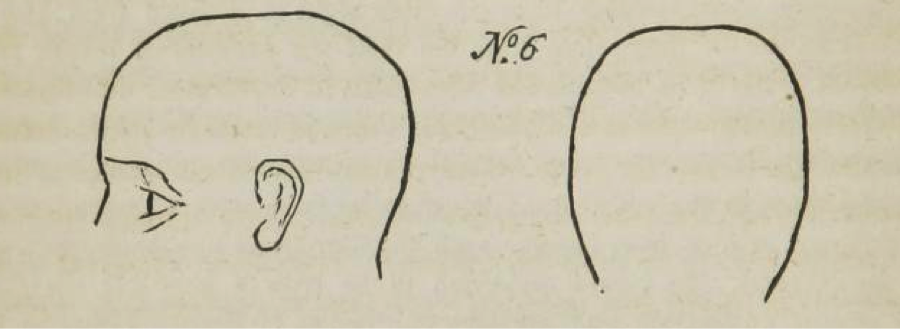
Figure 5: Farnham Cranial Outline No. 6
In stark contrast to No 6, Farnham asks readers-viewers to notice that the head of No 3 displays the “social characteristics of her race,” defined especially by “secretiveness and destructiveness”; Farnham’s verbal guide for her readers concludes with the assertion that No 3 is beyond reform, as “she will doubtless spend her life in prison, for she is constitutionally a criminal” (Rationale 9-10). Unlike No 6, the cranial outline of No 3 purports to show the biological and social inevitability of her character. As Farnham makes evident, the representational interaction she intends her readers-viewers to have with her images depends on them on carrying out her ideological program, one in which there are clear and apparent racial distinctions, which in turn are tied to inherent qualities or characteristics that distinguish women who can be reformed from women who are “constitutionally” criminal.
A similar racial logic is evident in Farnham’s description of the Brady daguerreotypes of women of color and immigrant women. The images of women, such as “T.Z….a Jewess of German birth and parentage,” show that “destructiveness is enormously developed, with large secretiveness…and very defective benevolence”; these “defective organs” reveal the inability to be reformed, even under Farnham’s “watchful eye” at Sing Sing (158-60). In Farnham’s visual-verbal narration, non-white women almost invariably lack the cranial capacity for reformation, and it would seem Farnham deploys phrenology as a way to excuse her own inability to rehabilitate certain prisoners.
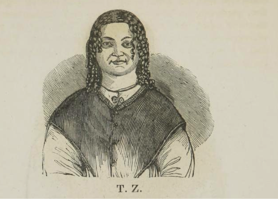
Figure 6: Farnham-Brady Daguerreotype: TZ
Under the guise of scientific knowledge, the visual rhetoric of phrenology enacted a process by which viewers could participate in the classification or marking of the criminal as criminal, and—as the Fowlers and Farnham did—use the visual mode as a means of social control. In this way, the process of phrenological “looking” and “seeing” is both ideological and surveillant, in the sense that Foucault intends. A reminder of Foucault’s ideas about the function of the medical examination in the nineteenth-century as a mode of disciplinary surveillance is useful here, particularly his definition of the “scientific gaze” as a way “to see everything” both inside and outside of the human body (173). Foucault’s focus on the emergence of technologies of observation such as telescopes, other lenses, and the examination practices of disciplinary institutions may be different than phrenology as a visual technology, but his emphasis on the ways that individuals were qualified and classified through a scientific “normalizing gaze” helps to explain how the visual rhetoric of phrenology worked similarly as an apparatus of power and control (DP 184). In short, the phrenological images used by Farnham exert this representational surveying power of “hierarchical observation” designed to establish a system of “visibility” in which criminal-subjects are presented as “objects…of the gaze” (DP 184-88).
Farnham’s phrenological sketches are not especially complex as drawings, and are not even particularly realistic (did all of these criminals have shaved heads that allowed their skulls to be traced?); similarly, the daguerreotypes do not seem to show anything especially notable in their portraiture (and in fact look comparable to Matthew Brady’s other daguerreotypes and photographs of people in this era—an interesting point, perhaps, to develop in another paper). Yet, Farnham asks her audience to view these images as tacit proof of scientific fact, begging the question: why might readers-viewers have believed or been persuaded? There have been considerable studies in cognitive science and neuroethics on why “we accord a special veridical status to photographs,” other images, and more recently to brain scans that appear to “invite us to believe,” as Keehner, et al. note. The findings of this literature conclude: “images with visual properties…may enhance feelings of directness and fluency, making naïve viewers more likely to be convinced by what they see” (426). In presenting her images as indices of objective realities, Farnham’s visual argument would seem to align with these contemporary findings insofar as she prompts a participatory experience on the part of her viewers, giving them the ability to corroborate and authorize their human vision as they gain fluency in this picturing process.
“Seeing is Believing”: The Persuasive Power of Brain Images
In a 2009 Scientific American article, neuroscientists Claus C. Hilgetag and Helen Barbas describe the brain’s cerebral cortex, filled with gray matter (“gelatinous tissue packed with neurons”), as “corrugated,” each brain cortex with “its own characteristic pattern of convolutions.” They liken these convolutions of the brain to an “intricate landscape of hills and valleys” folded into the skull, and point out that this uneven, grooved “topography” both gives the human brain its shape and holds the “network of nerve fibers” that “mediates our perceptions, thoughts, emotions and actions” (66-67). Notably, these contemporary research scientists also connect current scientific interest in neural discoveries and strategies for diagnosing and treating mental disorders to phrenology (Gall’s work in particular). Rather than dismissing phrenology altogether as a pseudoscience, Hilgetag and Barbas instead note its relevance—or at least resonance—to neurobiology: “evidence from novel brain-imaging techniques, aided by sophisticated computational analyses, has lent fresh support to some of those 19th-century notions” (66-67). Hilgetag and Barbas are among many scientists stating these connections and underscoring that an interest in understanding brain structure and trying to locate and isolate its different regions remains pervasive, with “noninvasive imaging techniques,” such as MRI, the scientific standard in ascertaining the origins of neurological diseases (68).
Hilgetag and Barbas also recognize that the use of neuroimaging extends beyond disease diagnostics in ways that suggest other direct connections to nineteenth-century practices: “modern neuroimaging methods have also enabled scientists to test the phrenological notion that cortical convolutions or the amount of gray matter in different brain regions can reveal a person’s talents” (70). In particular, the use of functional MRI (fMRI), which measures changes in blood flow and oxygenation and maps these changes through software that shows areas of increased neural activity as colored spots or areas in the brain’s gray matter, has emerged as a customary tool for imaging brain activity. The “map” currently in use for such technology digitizes areas of the brain in ways remarkably similar to the numbered phrenological head maps of the nineteenth-century.
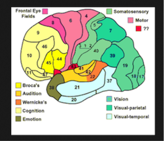
Figure 7: fMRI Numbered Head Map
As Walsh and Olman explain, the fMRI is a visual technique used to “trace patterns of neural activity in participants’ brains as they perform behaviors such as looking at pictures, reading text, and sometimes making decisions about what they are reading or viewing” (77). Many scientists claim that this four-dimensional picture of brain activity can be correlated with behaviors in participants, and the fMRI is often “employed rhetorically as evidence confirming or contesting” the hypotheses about such correlations (Walsh and Olman 79). There are numerous scientific studies using fMRI as a way to make a visual confirmation associated with a range of behaviors and inclinations, revealing conditions related to disorders such as epilepsy, autism spectrum disorders, and schizophrenia, or (by localizing areas associated with cognitive, emotional, and spiritual functions) showing what we are dreaming, predicting who we will love and marry, determining why we might or might not believe in God, and revealing whether we are lying.8
This last area of fMRI use in lie detection marks another connection to phrenology, especially to interests in criminal phrenology such as Farnham’s, highlighting a form of surveillant looking, to recall Cartwright, that turns part of the body “inside out” to reveal it to “public view” (xi). In the use of fMRI in lie detection, the medical gaze often functions also as a juridical one, especially in legal cases where image-scans decoding the patterns of brain activity are forms of evidence used to establish truths and delineate criminal from noncriminal actions and intents.9 In their 2014 study on the scientific, social, and ethical concerns of fMRI use for lie detection in criminal cases, Farah et al. note that “fMRI-based lie detection…has received the greatest public scrutiny” in recent years, in part because of attempts to admit these diagnostics as scientific evidence in a number of court cases ranging from trials for fraud to murder (128). There are numerous such studies by legal scholars and social and behavioral scientists examining this new area of neurolaw, such as the work by Saks et al. in which they note that “brain images are the most recent forms of evidence presented [in criminal trials] for the purpose of supporting factual arguments about the defendant’s mental condition” (105). The below example of fMRI imaging shows areas of the brain with higher concentrations of lies and truth, using red and blue accordingly, and in their review of the literature on the use of such images, Saks et al. conclude that “these images are inordinately persuasive” in convincing viewers that these images convey certain “facts” about a subject’s-defendant’s behavior (106).
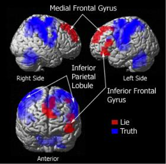
Figure 8: fMRI "Lies and Truth" Map
Farah et al. note that (as of 2014) there have been “at least three attempts to have fMRI-based lie detection admitted into US courts since 2010,” though the use of neurological images accompanying medical expert testimony is in widespread use. Although most of the studies using fMRI to detect lies have occurred in controlled laboratory environments, there continues to be interest in both medical sciences and the law in whether or not laboratory-derived indicators generalize to the real world. In particular, the use of fMRI in criminal cases has prompted much discussion and analysis of the ways that this visual form of forensic evidence seems “inordinately persuasive,” as Saks et al. remark about the effect of neuroimages on jury decisions in criminal trials.
Citing numerous studies, Saks et al. assert that the impact created by the use of brain scans “can lead…to unshakable conclusions”; even more than “interview-based diagnoses” by scientific and medical experts, or a “radiologist’s verbal description of [fMRI] findings… seeing the image of a brain with their own eyes leads judges and jurors to believe something that they will be unable to deny” (106). One such study, conducted and published in Cognition by psychologists David P. McCabe and Alan D. Castel, focused especially on the persuasive influence of neuroimages, and why “seeing is believing” in terms of “public perception of research on cognition” (343). McCabe and Castel note, importantly, that brain imaging research is controversial in terms of how fMRI data is interpreted by the lay public and in the media, particularly in the construction of a sort of “neuro-realism” that presents imaging phenomena as “uncritically real, objective…in the eyes of the public” (344).10 They conclude, nonetheless, that brain images may be persuasive “because they provide a tangible physical explanation for cognitive processes”—a form of “physical evidence” with a direct “appeal to people’s reductionist approach to understanding the mind as an extension of the brain” (349-50).
This emphasis on persuasion in the current scientific research on brain imaging further connects contemporary visual diagnostics and nineteenth-century visualizing techniques, such as phrenology. In her work analyzing the “transparent body as a social and cultural construct,” José van Dijck shows that since the nineteenth-century, “direct sensory perceptions” have been an “important diagnostic means” for translating the body into a “readable,” knowable visual text (5). In van Dijck’s sense, the body—or more specifically the head in this instance—is a “contested cultural object” that needs to be brought into view—and managed—through the “visual and representational instruments” and practices designed to observe, measure, diagnose, and allegedly treat the peculiarities of human physiology (4). Just as contemporary brain imaging provides a persuasive visual graphic for registering and disclosing causal relationships between the latent pathologies “beneath the skin,” as van Dijck says, and the outward manifestations of those pathologies, so, too, did Farnham’s phrenological images function rhetorically as persuasive visual mechanisms that rendered the head transparent and shaped viewers’ beliefs and normative ideals about the human body.
It might seem as though contemporary imaging lacks one important connection to nineteenth-century phrenological scanning: that DIY element. While most people who get a brain MRI are not guided by a radiologist or researcher through the steps on how to “read” it, are we so far off from having it within our power to do so? Just as jurors and judges are guided through how to interpret brain scans, and are persuaded by “seeing the image of a brain with their own eyes,” which leads them to “believe something that they are unable to deny,” so, too, might the lay public eventually learn to read heads—their own and others—and draw inferences from what they see (Saks et al. 106).
Conclusion
Just as there was considerable skepticism in the nineteenth-century about the promises of head reading as a means to diagnose and treat a range of mental disorders and diseases, there continues to be skepticism about brain imaging as a determinative technology, with some neuroscientists and science writers seeing the “statistical” claims about “subjects’ brains” as more phrenological “metaphor” than scientific “causal inference” (Shermer 46-48). Much of this skepticism, whether about phrenology or its new forms in neuroscience, stems from the sense that these scientific predictive promises are speculation; in neuroscience and the law, for example, this speculation is seen in the ways that scientific “promises elide a fundamental and perilous chasm between the aims and methods” of science and the uses of that science (Buckholtz R861). A similar “chasm” existed in the promises of phrenology, and it was this chasm that Farnham and other phrenologists attempted to bridge through their purposeful use of visual rhetoric.
As I have argued in this paper, Farnham’s use of phrenological images alongside her detailed descriptions of these images functioned rhetorically—and my response to these word-image texts has also been rhetorical. The derivatives of this visual rhetoric of phrenological heads are still with us especially in neuroimaging, demonstrating that the visual properties associated with head reading continue to be persuasive mechanisms for understanding human qualities and characteristics. While there may be skepticism about the promises of these forms of surveillant looking, there does seem to be general consensus that brain scans do show something, and that there are in fact “regions” of the brain responsible for our behaviors or processing of emotion—determinations not all that different than what Farnham and the phrenologists offered.
- 1. Farnham is notable for her work as a female prison reformer, serving as warden of the women’s section at Sing Sing long before a growing trend of feminist prison activists in the decades following the Civil War. For a history of women prison reformers in the nineteenth century, see especially Estelle B. Freedman.
- 2. These studies of the history of phrenology and other nineteenth century pseudosciences have been particularly useful in my work: Beer, G. , Cooter, R., Davies, J.D., Fabian, A., Hartley, L., Stern, M.B., Tomlinson, S. I have also benefited from the following work on Farnham and nineteenth-century prison reformers: Freedman, E., Lewis, W. D., Rafter, N.H.
- 3. Sampson’s book is one of many treatises in this era drawing connections between phrenology and early criminology, and even into the twentieth-century was viewed by some historians as “one of the best expositions…made by some nineteenth-century prison reformers to use phrenology” to explain criminal behaviors and propose methods to change them (Lewis vi).
- 4. My definition of ekphrasis aligns with contemporary critical views of visual rhetoric by W.J.T. Mitchell, Marguerite Helmers, Cara Finnegan, and Sonja K. Foss (among others, especially the contributors to the collection Defining Visual Rhetorics, edited by Helmers and Charles A. Hill). I am working with an understanding of ekphrasis through a visual rhetorical approach that goes beyond the interplay of word and image to consider a wider archive of images as “themselves carriers of meaning” (Helmers 64).
- 5. Nineteenth-century phrenology was influenced in part by the work of Johann Caspar Lavatar, a Swiss priest in the eighteenth-century, who sought to understand humanity by classifying human nature and character according to the anatomic forms of the head and face, and the outward signs of facial expression or physiognomy. According to Lavatar, the features of the head and face provided verification of otherwise hidden, internal truths about an individual, the essence or the soul that was mapped onto external appearance.
- 6. Another important figure in this history of phrenology from Gall in Europe to the Fowlers in the U.S. is George Combe, a Scottish lawyer turned phrenologist and the founder of the Edinburgh Phrenological Society. His most popular work, The Constitution of Man (1828), attracted practitioners in the U.S., such as the Fowlers, especially because of his application of phrenology to individual reform and social improvement.
- 7. While historians claim that Lombroso and other criminal anthropologists rejected phrenology as a valid means of determining anomalies and degenerative behaviors in criminal women, the visual history from Farnham to Lombroso suggests a different narrative. Most apparently, there are visual similarities in terms of the techniques employed to display and describe the features of female criminal heads, as seen in Lombroso’s drawing of an “anomalous brain of a [female] child murderer.” This image lifts techniques from phrenological diagrams of brains labeled to show distinct anatomical parts and functions. Lombroso’s use of photographs, too, while considered by many historians to be unique examples of visual evidence of how physiognomy proved delinquency and degeneration, can easily be traced back to Farnham’s use of the Brady daguerreotypes earlier in the century. Lombroso’s accompanying descriptions of the women in these photos, which include biographical “facts” linked to their physiognomic defects, also show a clear narrative trajectory from Farnham.
- 8. Most fMRI studies are performed on humans, though there are recent studies summarized in a 2013 Smithsonian article claiming to show the mental processes of dogs and their ability to think and feel in ways similar to humans.
- 9. In 2006, two companies began offering lie detection fMRI, No Lie MRI and Cephos, which claim that their technology can be used in a number of ways and circumstances, including business, family life, criminal justice, and national security.
- 10. There are numerous studies that point to the practical, scientific, and ethical concerns of using neuroimaging techniques, especially in relation to the law, all of which note the oversimplification of the data and tendency to misrepresent conclusions. See Bizzi et al, Using Imaging to Identify Deceit: Scientific and Ethical Questions, Farah et al, “Functional MRI-based Lie Detection: Scientific and Societal Challenges,” Buckholz and Faigman, “Promises, Promises for Neuroscience and the Law,” and Michael et al, “On the (Non)Persuasive Power of a Brain Image,” to list a few.
Beer, Gillian. Open Fields: Science in Cultural Encounter.Oxford, Oxford UP, 1996. Print.
Bittel, Carla.“Woman, Know Thyself: Producing and Using Phrenological Knowledge in 19th-Century America.” Centaurus, 55 (2013): 104-130. Print.
Bizzi, Emilio, et al. Using Imagine to Identify Deceit: Scientific and Ethical Questions. Cambridge: American Academy of Arts and Sciences, 2009. Print.
Buckholtz, Joshua W. and David L. Faigman. “Promises, Promises for Neuroscience and Law.” Current Biology 24:18 (2014), R861-R867. Print.
Burri, Regula Valerie. “Doing Distinctions: Boundary Work and Symbolic Capital in Radiology.” Social Studies of Science 38:1 (2008), 35-62. Print.
Cartwright, Lisa. Screening the Body: Tracing Medicine’s Visual Culture. Minneapolis: U of Minnesota P, 1995. Print.
Colbert, Charles. A Measure of Perfection: Phrenology and the Fine Arts in America. Chapel Hill: U of North Carolina P, 1997. Print.
Cooter, Roger. The Cultural Meaning of Popular Science: Phrenology and the Organization of Consent in Nineteenth-Century Britain.Cambridge: Cambridge UP, 1984.Print.
Davies, John Dunn. Phrenology, Fad and Science: A 19th Century American Crusade. 62. New Haven: Yale UP, 1955. Print.
Fabian, Ann. The Skull Collectors: Race, Science, and America’s Unburied Dead. Chicago: U Chicago P, 2010. Print.
Farah, Martha J. et al. “Functional MRI-based Lie Detection: Scientific and Societal Challenges.” Nature 15 (February 2014), 123-131. Print.
Farnham, Eliza W. Woman and Her Era. New York: A.J. Davis & Co., 1864. Print.
---. "Notes and Illustrations." In Rationale of Crime and its Appropriate Treatment, Marmaduke Sampson. (1846) Reprinted Edition by Patterson Smith Publishing, Montclair, NJ, 1973. Print.
Fee, Elizabeth. “Nineteenth-Century Craniology: The Study of the Female Skull.” Bulletin of the History of Medicine, 53 (1979): 415-433. Print.
Finnegan, Cara A. “Doing Rhetorical History of the Visual: The Photograph and the Archive.” In Defining Visual Rhetorics, eds. Hill and Helmers. Mahwah: New Jersey: Lawrence Erlbaum Associates, Inc. Publishers (2004); 195-214. Print.
Foss, Sonja K. “Framing the Study of Visual Rhetoric: Toward a Transformation of Rhetorical Theory.” In Defining Visual Rhetorics, eds. Hill and Helmers. Mahwah: New Jersey: Lawrence Erlbaum Associates, Inc. Publishers (2004); 303-314. Print.
Foucault, Michel. Discipline and Punish: the Birth of the Prison. Trans. Alan Sheridan. New York: Vintage Books, 1977. Print.
Fowler, O.S. and L.S and Samuel Wells. The American Phrenological Journal. New York: Fowler and Wells Co., Publishers, 1838-1911. Print.
Fowler, O.S., and Fowler, L.N. The Illustrated Self-Instructor in Phrenology and Physiology. New York: Fowlers and Wells, 1849. Print.
Freedman, Estelle B. Their Sisters’ Keepers: Women’s Prison Reform in America, 1830-1930. Ann Arbor: U Michigan P, 1981. Print.
Goggin, Maureen Daly. “Visual Rhetoric in Pens of Steel and Inks of Silk: Challenging the Great Visual/Verbal Divide.” In Defining Visual Rhetorics. Eds. Charles A. Hill and Marguerite Helmers. Mahwah, NJ: Lawrence Erlbaum Associates, 2004. 1812- of 6169. Ebook.
Gries, Laurie E. “Emerging Methods of Visual Rhetorics.” JAC 29:1/2 (2009), 437-450. Web.
Hartley, Lucy. "A Science of Beauty? Femininity, Fitness and the Nineteenth-century Physiognomic." Women: A Cultural Review 12.1 (2001): 19-34. Print.
Helmers, Marguerite. “Framing the Fine Arts Through Rhetoric.” In Defining Visual Rhetorics, eds. Hill and Helmers. Mahwah: New Jersey: Lawrence Erlbaum Associates, Inc. Publishers (2004); 63-86. Print.
Hilgetag, Claus C. and Helen Barbas. “Sculpting the Brain.” Scientific American 300:2 (Feb 2009), 66-71. Print.
Hill, Charles A., and Helmers, Marguerite, eds. In Defining Visual Rhetorics. Mahwah, NJ: Lawrence Erlbaum Associates, Inc. Publishers, 2004. Print.
Keehner, Madeleine, et al. “Different Clues From Different Views: The Role of Image Format in Public Perceptions of Neuroimaging Results.” Pynchon Bull Rev 18 (2011), 422-428. Print.
Lewis, W. David. From Newgate to Dannemora: the Rise of the Penitentiary in New York, 1796-1848. Ithaca: Cornell UP, 1965. Print.
McCabe, David P. and Alan D. Castel. “Seeing is Believing: The Effect of Brain Images on Judgments of Scientific Reasoning.” Cognition 107 (2008), 343-352. Print.
Michael, Robert B., et al. “On the (non)Persuasive Power of a Brain Image.” Pynchon Bull Rev 20 (2013), 720-725. Print.
Mitchell, WJT. Picture Theory. Chicago: U Chicago P, 1994. Print.
Rafter, Nicole Hahn. Partial Justice: Women in State Prisons, 1800-1935. Boston: Northeastern UP, 1985. Print.
Rafter, Nicole Hahn, and Gibson, Mary, eds. Criminal Woman, the Prostitute, and the Normal Woman: Cesare Lombroso and Guglielmo Ferrero. Durham: Duke UP, 2004. Print.
Saks, Michael J. et al. “The Impact of Neuroimages in the Sentencing Phase of Capital Trials.” Journal of Empirical Legal Studies. 11:1 (March 2014), 105-131. Print.
Sekula, Allan. “The Body and the Archive.” October 39 (Winter 1986): 3-64.
Shermer, Michael. “A New Phrenology?” Scientific American 298:5 (May 2008), 46-48. Print.
Snyder, Joel. “Visualization and Visbility.” In Picturing Science, Producing Art, eds. Caroline A. Jones and Peter Galison. New York: Routledge (1998), 379-400. Print.
Stern, Madeline B. Heads and Headlines: the Phrenological Fowlers. Norman: U of Oklahoma P, 1971. Print.
Tomlinson, S. Head Masters: Phrenology, Secular Education, and Nineteenth-Century Social Thought. Tuscaloosa: U of Alabama P, 2005. Print.
van Dijck, Jos. The Transparent Body: A Cultural Analysis of Medical Imaging. Seattle, WA: U of Washington P, 2005. Print.
Walsh, Lynda and Cheryl Olman. “Recommended Approaches to the Neuroimaging Literature on Autism Spectrum Disorders.” In Autism Spectrum Disorders in the College Composition Classroom, eds Val Gerstle and Lynda Walsh. Milwaukee, WI: Marquette UP (2011), 75-86. Print.